

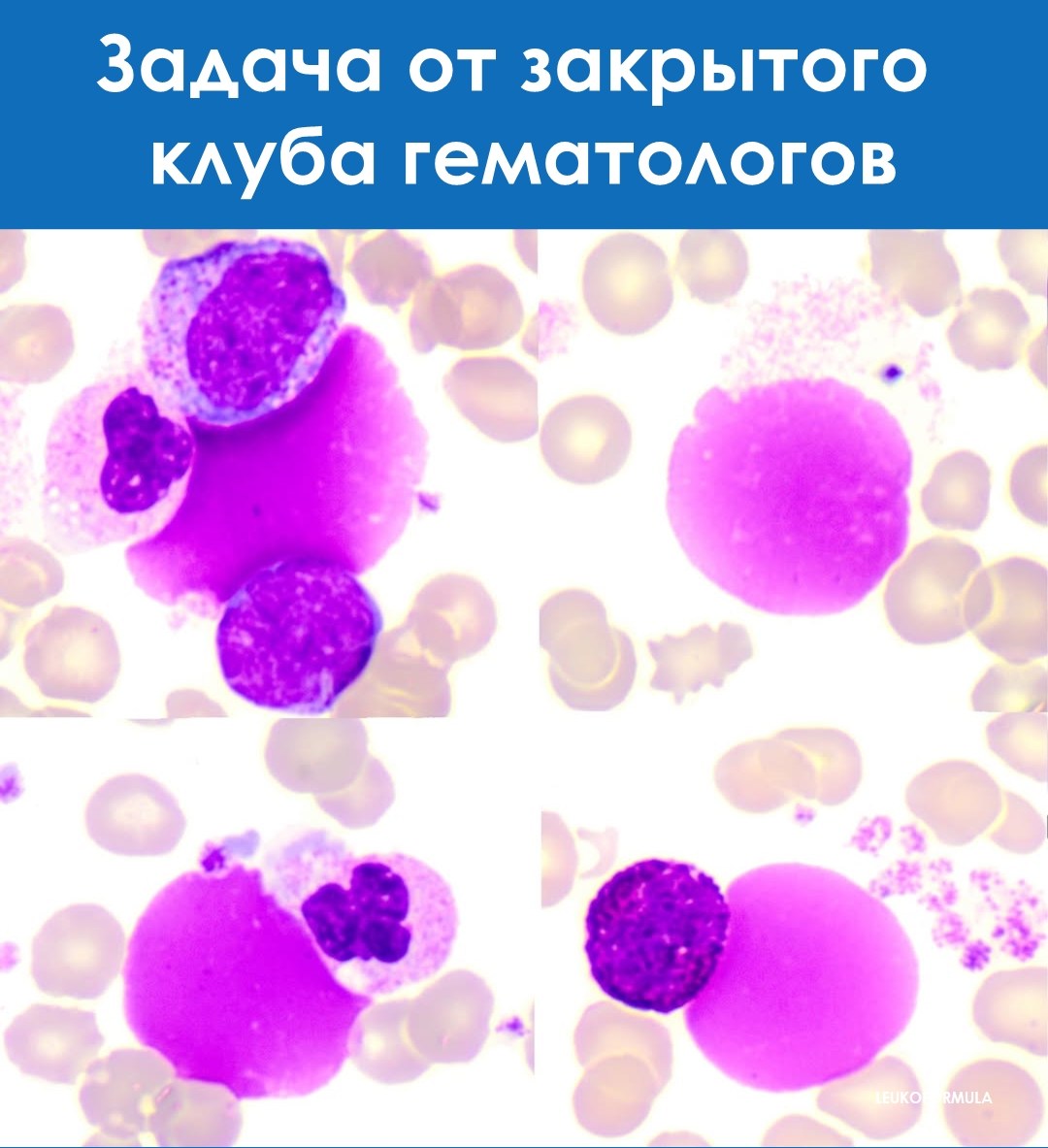
💡 Exercise from the Closed Club of Laboratory Hematologists. 🔬 Question. In chronic myeloid leukemia (CML), what do these “pink masses” in the blood smear represent? Choose one answer option: 🔍 Answer options: A – Artifacts B – Myeloid nuclear shadows C – Anucleated blasts D – Giant platelets E – New cytomorphological finding F – Cryoglobulin
The first 3 people who answer correctly in the comments will receive an invitation to the closed club. CORRECT ANSWER TOMORROW!


