

Parasitology Exercise
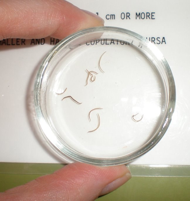
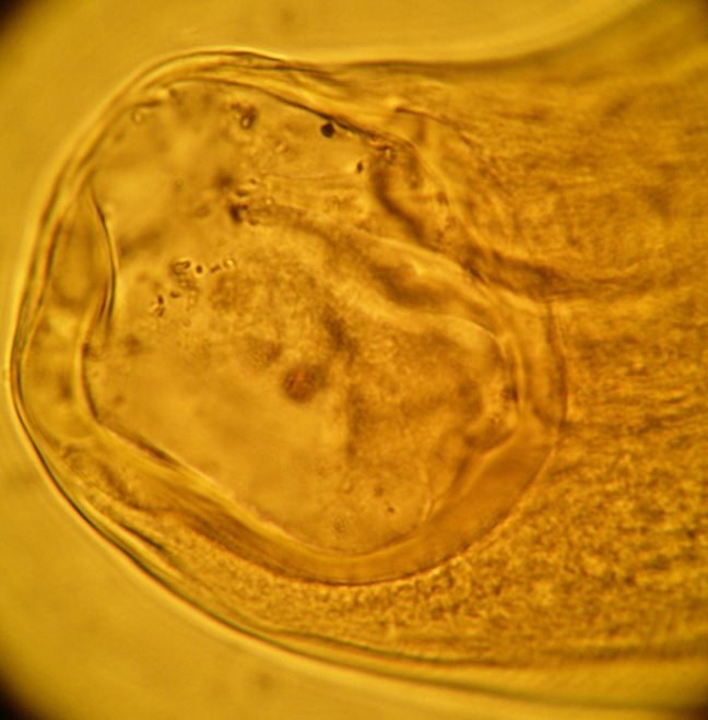
The following worms were removed from the small intestine during colonoscopy (image 1). During the procedure, the endoscopist noted several punctate hemorrhages in the surrounding mucosa and correctly concluded that these represented previous attachment sites of the worms. Light microscopy of the oral parts revealed the following (image 2). Can you identify the parasite? ANSWER TOMORROW!


