

🔬Clinical parasitology exercise (high difficulty!).
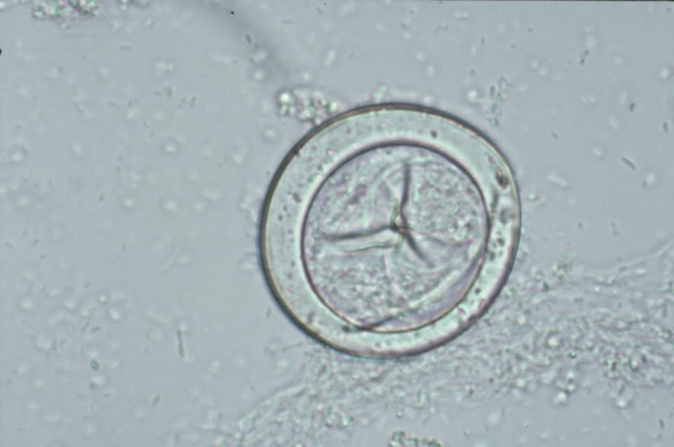
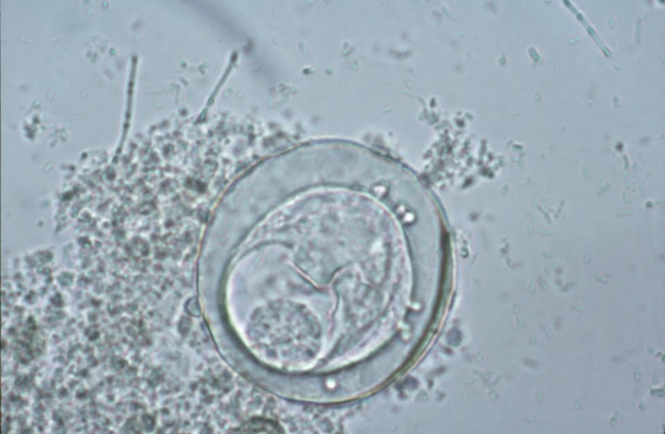
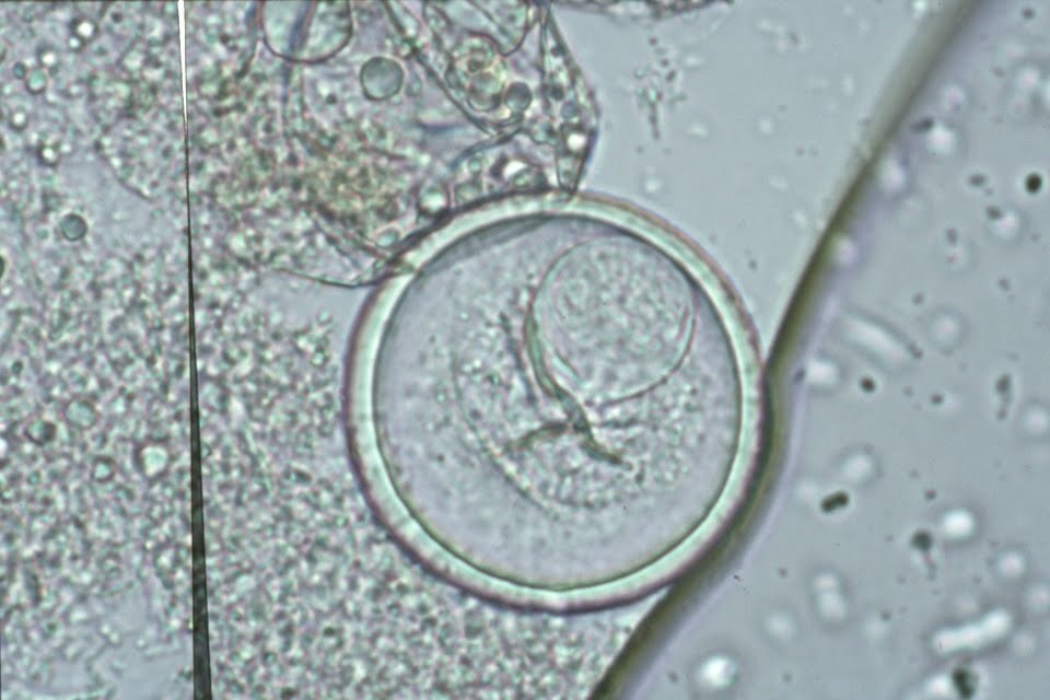
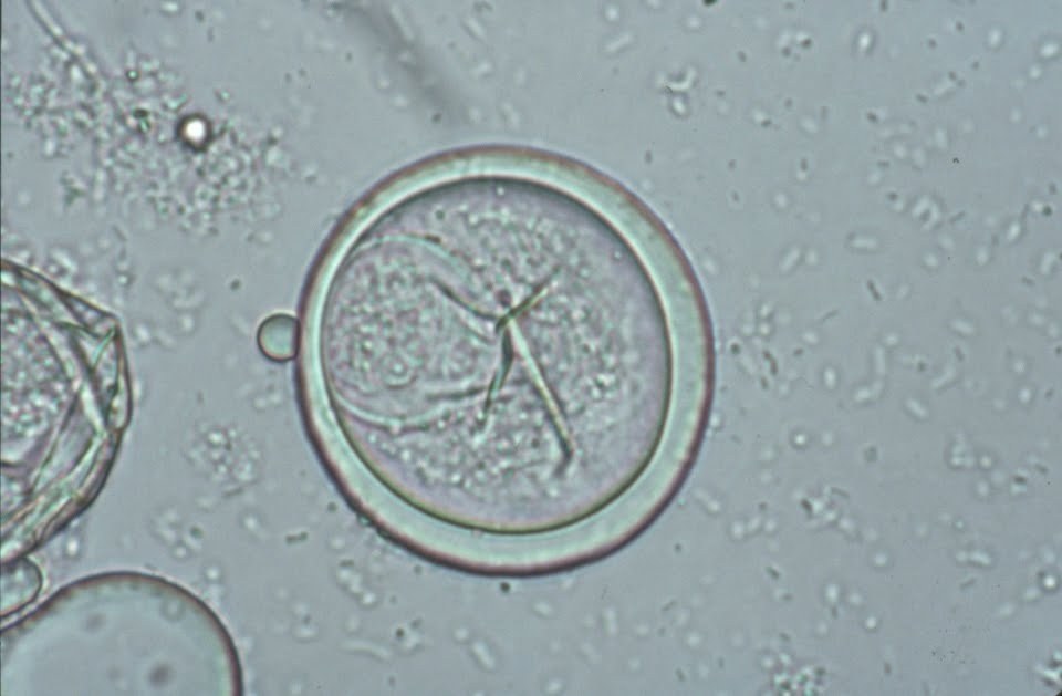
✅The following findings in the images were discovered in the stool of a patient working with animals and in contact with primates.
✅The objects shown have a diameter of about 40 microns. Unstained preparation, initial magnification x400.
❗ANSWER TOMORROW in this group.


