

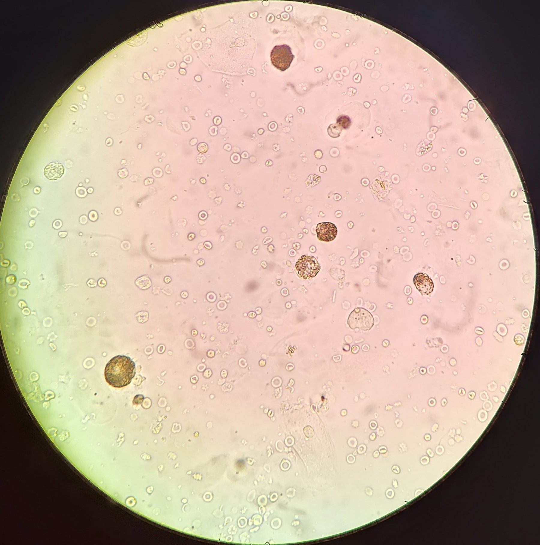
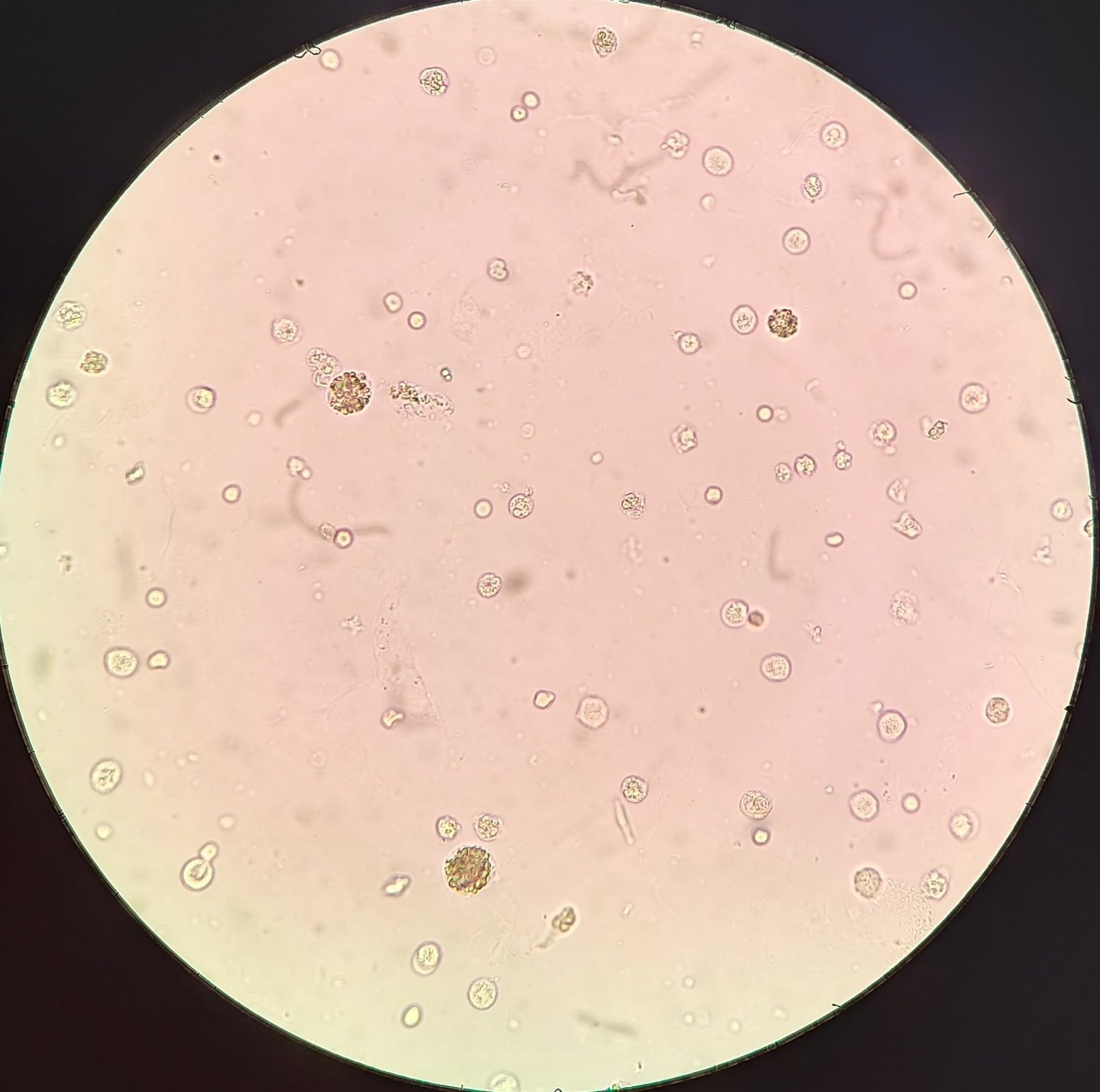
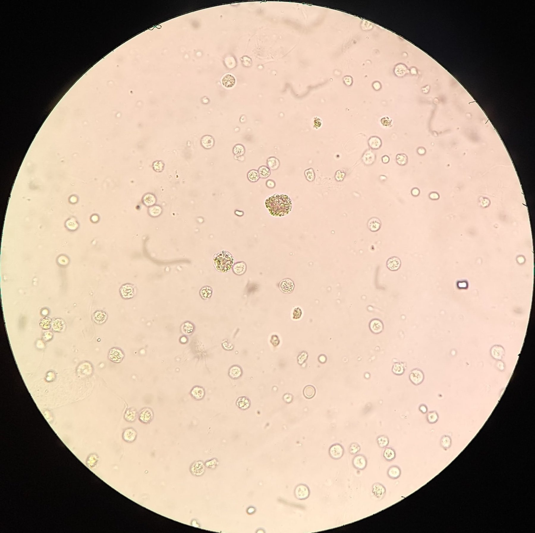
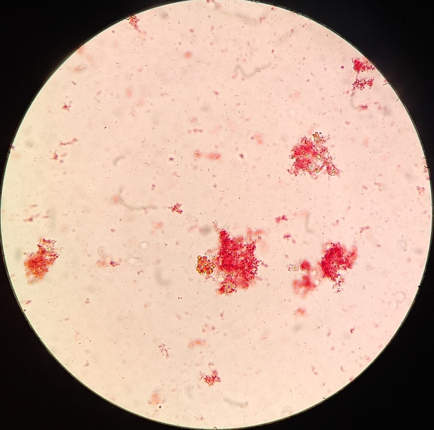
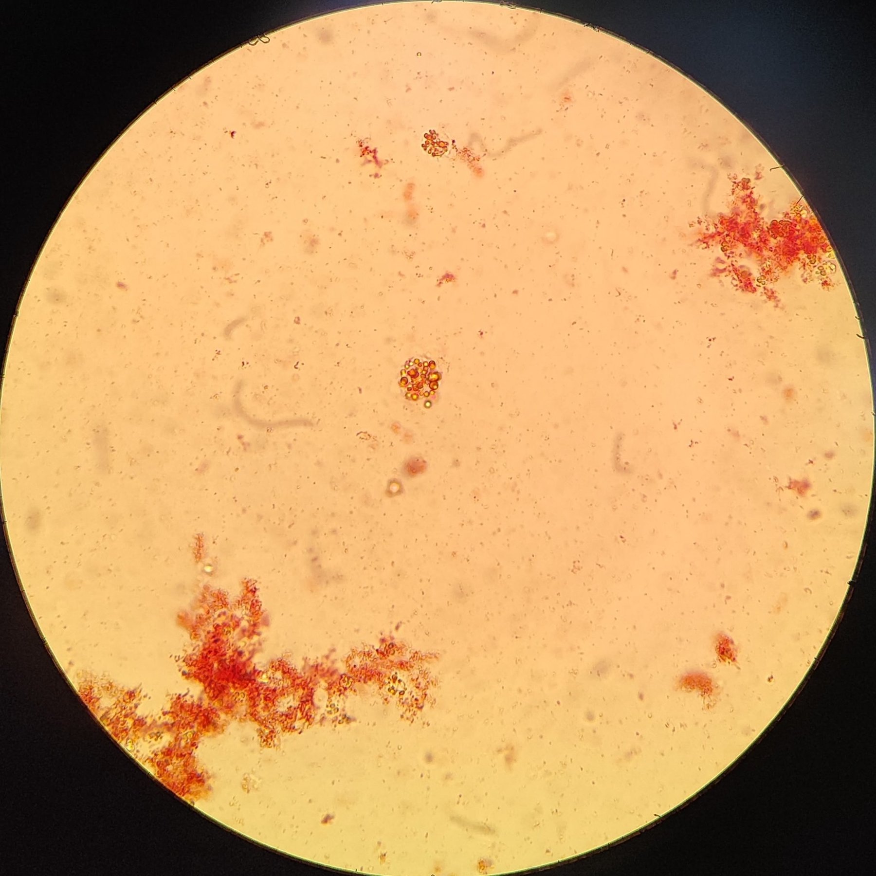
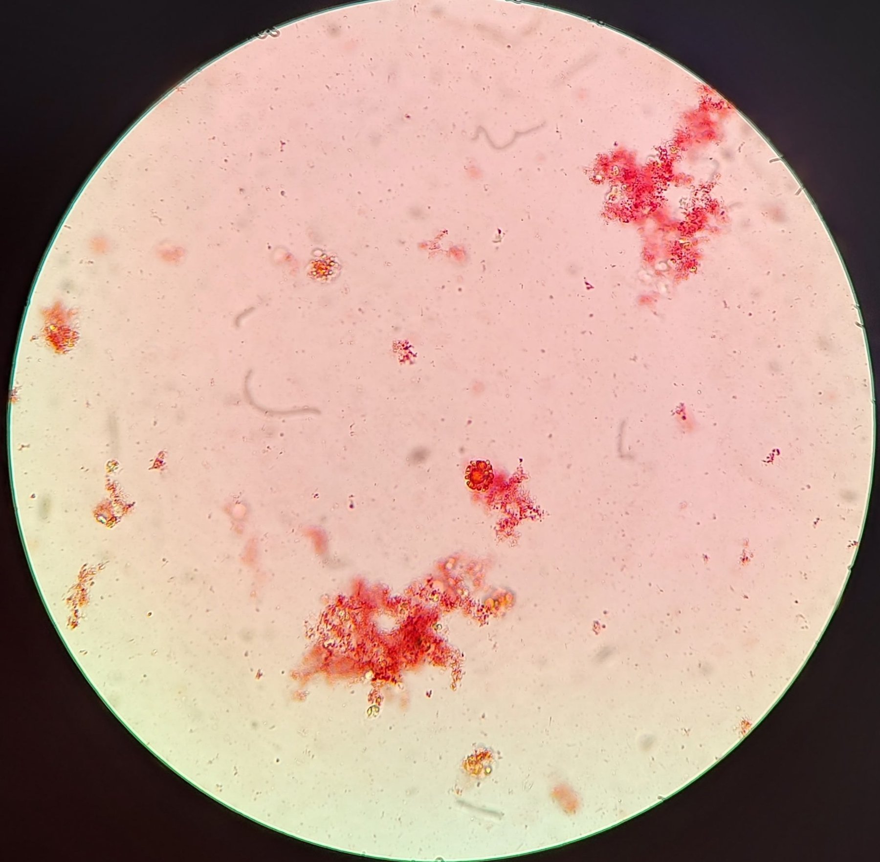
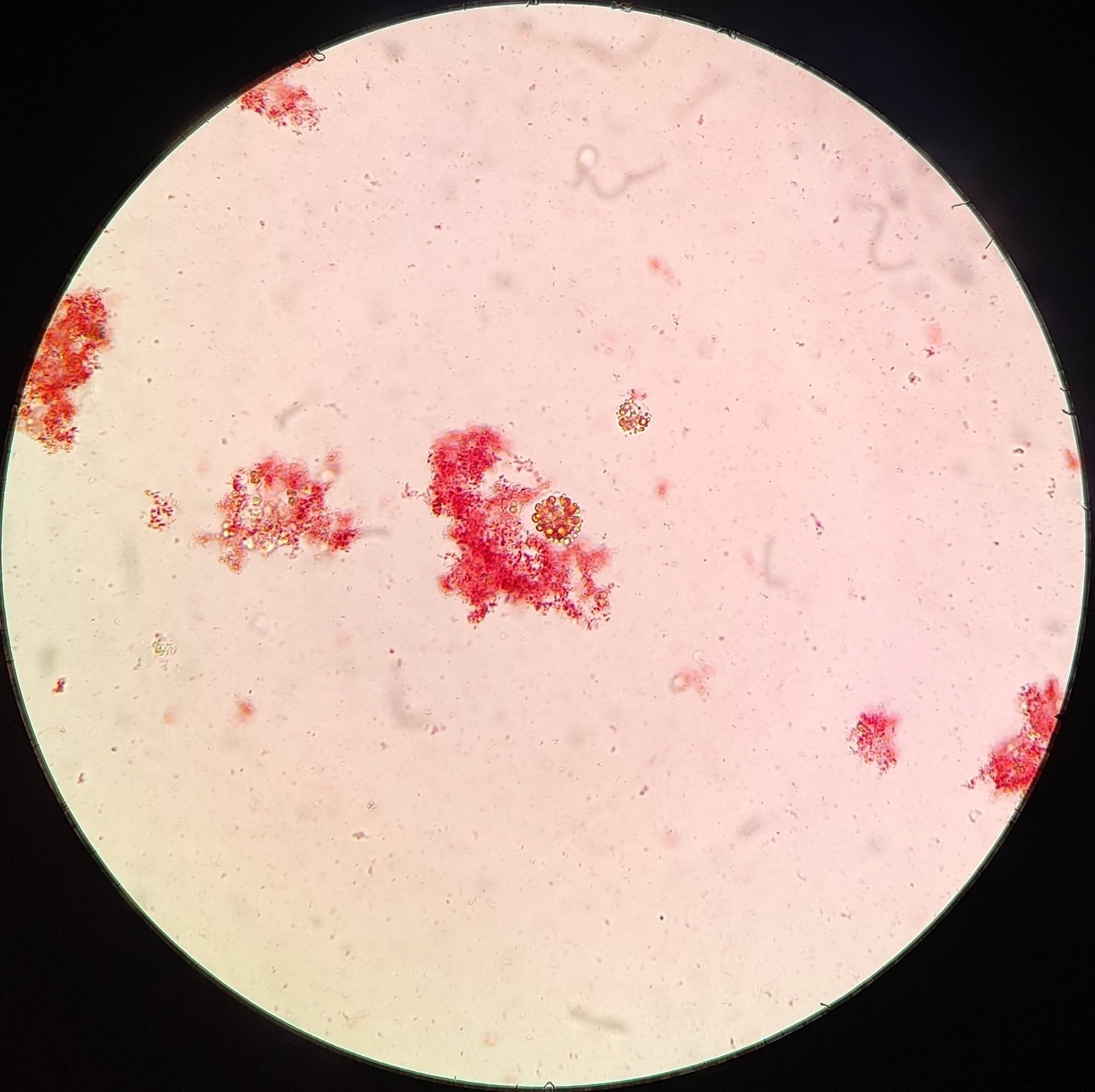
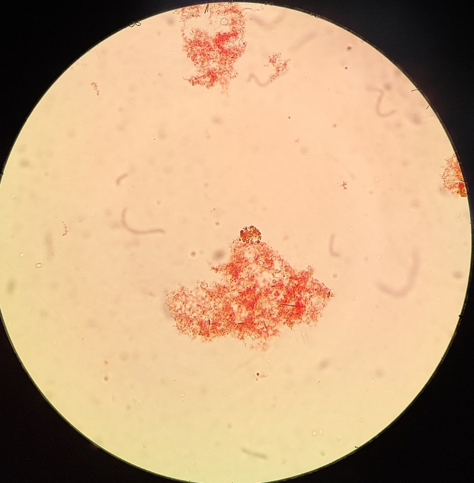
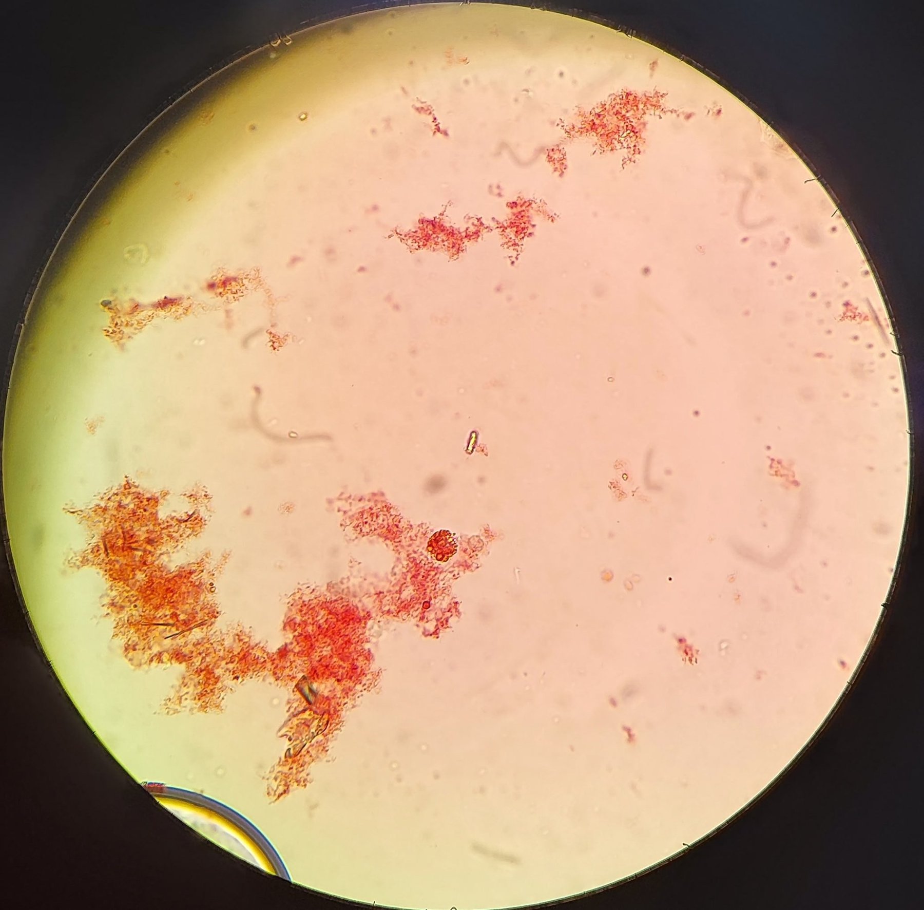
Renal epithelium with fatty degeneration. 1-3 photos - native preparation. Large cells packed with highly light-refractive granules of various sizes. 4-9 photos - preparation stained with Sudan III (red) and heated over an alcohol lamp flame. Since Sudan stains fatty elements, all brown granules changed color to yellow, orange, or red. Native and stained preparations, 400x magnification.
🙏 Material kindly provided by biochemist Daniil Adamov, Moscow


